Tumark® Vision Atlas
Case 2
Invasive ductal carcinoma in a 49 year-old patient
Prof. Dr. Katja C. Siegmann-Luz, Diagnostisches Brustzentrum Königs Wusterhausen, Germany
Case description
The patient presented with a new, three week old palpable finding in the right breast. Normal findings in the screening of dense glandular tissue and for macromastia. Ultrasound shows a malignoma-typical lesion, 24mm in diameter, in the top right at 12 o’clock, BI-RADS 5. Poorly differentiated triple-negative breast cancer NST G3 confirmed by ultrasound-guided punch biopsy. The lesion is marked with Tumark Vision (figure 1) with ultrasound guidance prior to neoadjuvant chemotherapy. A tumor, 26mm in diameter, surrounding the clip is differentiated in post-intervention breast tomosynthesis (figure 2).
Course of treatment
Ultrasound check-up after six months of neoadjuvant therapy shows partial remission of the tumor and an unchanged position of Tumark Vision (figure 3). Ultrasound-guided, pre-operative wire marking of the clip (figure 4), clearly visible in the segment resectate (figure 5). Tissue margins are tumor-free (R0).
Conclusion
Long-term ultrasound visibility of the placed clip is helpful in planned breast-conserving surgery (BCS) after neoadjuvant therapy, especially in the case of tumor remissions and if distinguishing of the tumor is limited. Thanks to its geometry, Tumark Vision can be easily distinguished as a hyperechoic ring-shaped structure.
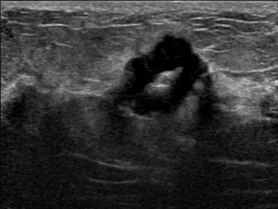
Case 2, fig. 1
Ultrasound depiction of Tumark Vision within the cancerous lesion measuring 24 mm prior to the planned neoadjuvant therapy.
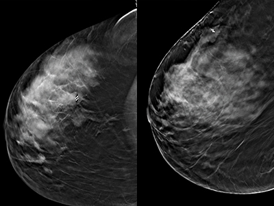
Case 2, fig. 2
Breast tomosynthesis right CC (left) and right ML (right), showing the clip (Tumark Vision) within the cancerous lesion measuring 26mm at the right top.
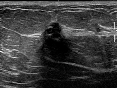
Case 2, fig. 3
Breast ultrasound after six months of neoadjuvant chemotherapy, showing Tumark Vision within the residual cancer, measuring 10mm after partial remission.
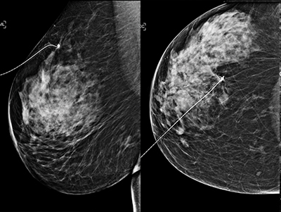
Case 2, fig. 4
Mammography right ML (left) and CC (right) images to monitor wire positioning after ultrasound-guided marking of Tumark Vision.
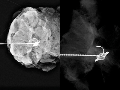
Case 2, fig. 5
Sample radiography. Diagnostic exposure (left), magnification of the raw image to show Tumark Vision (right).