Tumark® Vision Atlas
Case 1
47 year-old patient with cancer in the right breast
Prof. Dr. Katja C. Siegmann-Luz, Diagnostisches Brustzentrum Königs Wusterhausen, Deutschland
Case description
A tomosynthesis (figure 1) of a 47 year-old patient without clinical symptoms shows a lesion with surrounding architectural distortion. The ultrasound shows a correlating hypoechoic lesion. After an ultrasound-guided punch biopsy was performed (figure 2), a clip marker was positioned with Tumark Vision. Histological finding – Infiltrate of a moderately differentiated, invasive cancer of the ductal type/NST, as well as a small-lesion lobular neoplasia (LIN II). Tumor biology – Estrogen receptor: positive (100%); Progestin receptor: positive (100%); HER2/neu: moderate expression (score 2+). FISH negative.
Course of treatment
A pre-operative MRI shows the susceptibility artifact due to the clip ( 3). A segment excision (SE) was performed following the pre-operative ultrasound-guided wire marking (figure 4). Sample radiography (figure 5).
Conclusion
Good ultrasound visibility of the expanded clip in the hypoechoic tumor. Also, depiction of the mammographic and MR tomographic clip properties. Successful resection following pre-operative ultrasound-guided fine needle marking (FNM) of the tumor/clips.
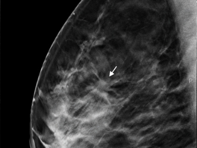
Case 1, fig. 1
Tomosynthesis-level image shows lesion with surrounding architectural distortion.
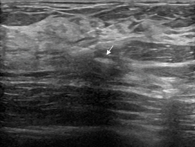
Case 1, fig. 2
Breast cancer shown as a hypoechoic lesion and the clip within (not yet expanded), directly after the procedure.
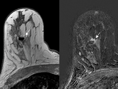
Case 1, fig. 3
Susceptibility artifact due to the clip in the T1-w dynamic subtraction. Examination performed approximately two weeks after clip procedure and a few days pre-surgery.
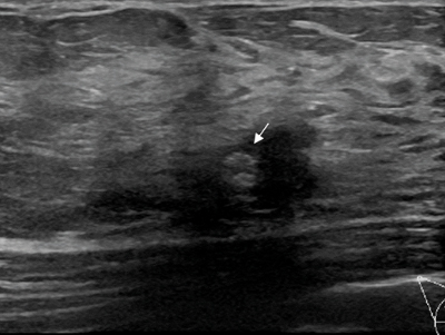
Case 1, fig. 4
Good ultrasound visibility of the expanded clip in the hypoechoic tumor prior to FNM.
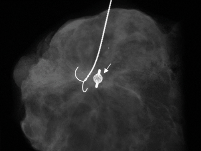
Case 1, fig. 5
Mammographic depiction of the marker.