Tumark® Vision Atlas
Case 4
A 37 year-old patient with invasive ductal cancer
Prof. Dr. Joerg Heil, Universitäts-Frauenklinik Heidelberg, Germany
Case description
The patient presented in February 2017 for an evaluation of an unclear finding in her left breast that was discovered during a screening session. Mammographic evidence of a lesion was found in two levels in the imaging, on the left side at 1 o’clock, extending to over 11 x 10mm in size, ACR 3, BIRADS 5. Ultrasound evidence of an unclear and suspicious lesion on the left at 1 o’clock position, extending to over 10 x 10 x 11 mm in size, BIRADS 5. An ultrasound-guided punch biopsy result is a moderately differentiated HER2 phenotype breast cancer NST G2. Prior to neoadjuvant chemotherapy, ultrasound-guided lesion marking was performed with Tumark Vision (figure 1).
Course of treatment
During ultrasound and mammography follow-ups carried out at two, three and five months after marking, the clipmarker was clearly visible in the ultrasound and mammographic imaging within the tumor bed (figures 2-6) in a finding no longer clearly distinguishable.
Upon completion of NACT consisting of 6 x TCbHP, segment resection with ALND was performed. After sample radiography and radiological examination (confirmation with a mammography), a second resection, medio-cranial, was recommended and performed. In sample radiography, the Tumark Vision was clearly visible in the resected segment tissue (figure 8). The histological finding suggests a pathological complete remission (ypT0, ypNo, Lo).
Conclusion
In the case presented, the Tumark Vision was clearly distinguished in the ultrasound scans as a hyperechoic, ring-shaped structure throughout the complete course of neoadjuvant therapy. Due to the limited tumor differentiation, here in complete remission already after the first NACT cycle, long-term dstinguishing of the clip was helpful during follow-ups.
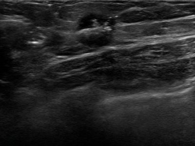
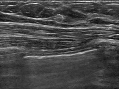
Case 4, fig. 1
Ultrasound depiction of the Tumark Vision within the cancerous lesion immediately after marking and before NACT initiation.
Case 4, fig. 2
Ultrasound depiction of the Tumark Vision two months after marking and after two NACT cycles. Finding cannot be clearly differentiated (anymore). Marker within the tumor bed. .
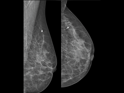
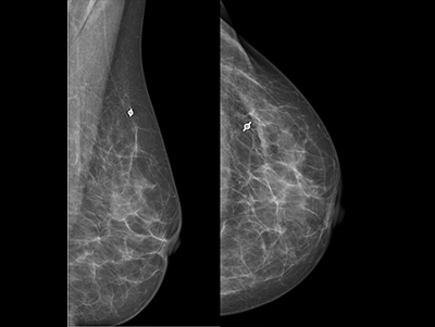
Case 4, fig. 3
Mammography left ML (left image) and CC (right image) two months after marking following two NACT cycles. Finding cannot be clearly differentiated (anymore), marker within the tumor bed.
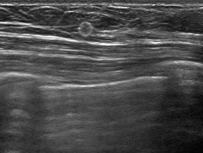
Case 4, fig. 4
Ultrasound depiction of the Tumark Vision three months after marking. Finding cannot be clearly differentiated (anymore), marker within the tumor bed.
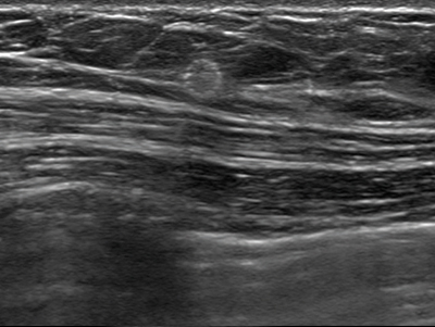
Case 4, fig. 5
Ultrasound depiction of the Tumark Vision five months after marking upon completion of NACT (6 x TCbHP). Finding cannot be clearly differentiated (anymore), marker within the tumor bed.
Case 4, fig. 6
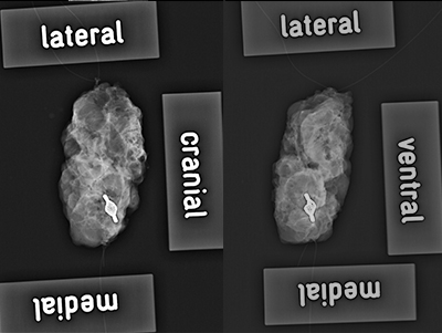
Case 4, fig. 7
Sample radiography